Study can advance the development of therapies for brain regeneration
Strokes lead to irreversible damage to the brain and are one of the most common causes of dependency or death. As the cellular reactions to a cerebral infarction are not yet fully understood, there is a lack of possible approaches to promote the regeneration of damaged nerve tissue in the brain. A study led by MedUni Vienna and recently published in “Nature Communications” closes crucial gaps in our knowledge and paves the way for research into new, targeted therapeutic strategies.
In order to investigate the activity of individual cells in the brain after a stroke, the research team led by first author Daniel Bormann (Department of Thoracic Surgery) and study leaders Hendrik J. Ankersmit (Department of Thoracic Surgery) and Michael Mildner (Department of Dermatology) used single-cell RNA sequencing in animal models, which are already established in stroke research. Using this method, the researchers were able to identify different cell types and their reactions in the early phase after a cerebral infarction. They focussed on two specific cell types (astrocytes and oligodentrocytes) from the group of glial cells, which are involved in many fundamental processes in the brain.
snRNAseq reveals differential cell cluster abundance and cluster-specifictranscriptional perturbations 48 h after ischemic stroke
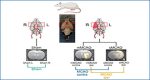
a Illustration of study design, depicting brain regions sampled for snRNAseq, from n = 4 Sham control rats and n = 7 middle cerebral artery occlusion (MCAO) group rats. MRI images of brain tissue from Sham-operated, moderate (mMCAO) and severe (sMCAO) MCAO group rats are presented, ischemic lesions are highlighted in orange. Rat schematic created with BioRender.com. MCAO ipsi MCAO group, hemisphere ipsilateral to ischemic lesion, MCAO contra MCAO group, hemisphere contralateral to ischemic lesion. b Uniform Manifold Approximation and Projection (UMAP) plot depicting 68616 nuclei annotated to 29 major cell clusters in the integrated dataset. Cell cluster AC astrocyte cluster, CHOL_IN cholinergic interneurons, EP_M_C ependymal and mural cell cluster, GABA_Amb Ambiguous GABAergic neuronal cluster, GABA_IN_Adarb2 + , GABA_IN_Adarb2- GABAergic interneurons, Adarb2 positive/negative, respectively, GABA_MSN GABAergic medium spiny neurons, GLU_Satb2 + , GLU_Satb2- Glutamatergic neurons, Satb2 positive/negative, respectively, OLIGO_1 immature oligodendrocyte lineage cluster, OLIGO_2 myelinating and mature oligodendrocyte lineage cluster. c–e Dotplots depicting curated marker genes for all major cell clusters. The dendrogram in (c) represents overarching taxons of identified major cell clusters. The dotplot in (d) depicts curated cluster markers of glutamatergic neurons. Colored bars next to the gene names denote established associations to cortical layers. Representative corresponding RNA in situ hybridization (ISH) results are depicted next to the colored bars. All RNA ISH studies were taken from Allen Brain Atlas database, and are referenced in detail in Supplementary Tab. 1. L = layer, CLA = claustrum, ec = external capsule, LSr = lateral septal nucleus, PIR = piriform cortex. e Dotplot depicting marker gene expression in cholinergic and GABAergic neurons. f Stacked bar plot depicting the relative abundance of each cell cluster within each sample. g Nuclei distribution coloured by treatment group. h Gene module score derived from the stroke-associated myeloid cell (SAMC) gene set13. i Strip plots depicting distribution of DEGs derived from MCAO ipsi vs Sham and MCAO ipsi vs MCAO contra comparisons, for all major cell clusters. DEGs were calculated using the MAST statistical framework as specified within the Methods section. Source data are provided within Supplementary data.
Cellular interaction in the infarct area
Scientists already know that astrocytes divide rapidly after a stroke and accumulate around the infarct area.
“We have been able to show, that oligodentrocyte precursor cells also divide in the acute infarct phase and accumulate at the edge of the damaged nerve tissue,” reports Daniel Bormann. In further complex analyses, the scientists discovered possible mechanisms in the wound healing of the damaged brain area: “We found considerable overlaps in the gene activity patterns of both cell types, especially in those genes that are important for building the glial barrier around the infarct,” Bormann explains a remarkable detail from the study.
In addition, it was discovered that certain immune cells (microglia and macrophages), which are also found in the vicinity of the infarct, release a signalling molecule (osteopontin). This molecule could play a decisive role in directing the glial cells to the edge of the infarct, where they are needed, among other functions, to form a barrier between the damaged and healthy tissue.
“Our description of the interaction between immune and glial cells in the infarct area contributes considerably to a better understanding of the regeneration processes of nerve tissue in the brain after a stroke,” says Daniel Bormann, summarising the significance of the results.
After cardiovascular diseases and cancer, strokes are the third most common cause of death in Austria and the most frequent cause of permanent disability and the need for long-term care. An ischaemic stroke is caused by the blockage of a blood vessel in the brain. The resulting interruption in the supply of oxygen and nutrients leads to a cerebral infarction in which nerve tissue is irreversibly damaged. Treatment approaches to date focus on restoring blood flow, which has to happen within 24 hours and is only possible for part of those affected. As the cellular processes following a cerebral infarction are not yet fully understood, treatment approaches to promote the regeneration of nerve tissue after a stroke are still lacking.
“The insights gained in our international and interdisciplinary study are a promising resource for developing novel targeted therapeutic strategies,” says Daniel Bormann with a view to further scientific investigations.
Source – Medical University Vienna
Bormann D, Knoflach M, Poreba E, Riedl CJ, Testa G, Orset C, Levilly A, Cottereau A, Jauk P, Hametner S, Stranzl N, Golabi B, Copic D, Klas K, Direder M, Kühtreiber H, Salek M, Zur Nedden S, Baier-Bitterlich G, Kiechl S, Haider C, Endmayr V, Höftberger R, Ankersmit HJ, Mildner M. (2024) Single-nucleus RNA sequencing reveals glial cell type-specific responses to ischemic stroke in male rodents. Nat Commun 15(1):6232. [article]