In the ever-evolving field of cancer research, scientists are continuously developing new ways to study how tumors interact with their surrounding environment, known as the tumor microenvironment (TME). One of the latest advancements in this area is a technique called spatial transcriptomics (ST). This cutting-edge method allows researchers to map out where genes are active within different regions of a tissue, giving them a detailed view of how cells within a tumor communicate and behave. However, despite the promise of ST, many existing tools that analyze this data have some limitations. They often overlook important tissue features, like the physical structure of cells, and rely too heavily on data from single-cell RNA sequencing, which doesn’t always capture the complexity of the TME.
To address these challenges, researchers at the University of Texas MD Anderson Cancer Center, have developed a new framework called the Morphology-Enhanced Spatial Transcriptome Analysis Integrator, or METI for short. METI is designed to provide a more comprehensive analysis of the TME by integrating three key components: spatial transcriptomics data, cell morphology (how cells look and are structured), and curated gene signatures (specific patterns of gene activity associated with particular cell types or states).
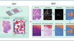
Workflow of METI
METI takes 10x Visium Spatial Transcriptomics (ST) data, with a spot-by-gene matrix for gene expression data, Hematoxylin and Eosin (H&E) images, and XY coordinates that map the location of each spot onto the image as input. With METI algorithm, METI offers cell type identification, nuclei segmentation and the functionality of generating 3D cell density plots in five distinct modules. Module 1 is dedicated to mapping normal and premalignant cells through the integration of gene expression (GE) data and H&E images. Module 2 focuses on identifying cancer cell domains and characterizing their heterogeneity. Module 3 is dedicated to T cell mapping and phenotyping. Module 4 involves in-depth analysis of other immune cells. Lastly, Module 5 pertains to the analysis of Cancer-Associated Fibroblasts (CAFs).
So, how does METI improve our understanding of cancer? First, it helps researchers map out where different types of cancer cells and other cells in the TME are located within the tissue. This is important because the physical location of these cells can influence how they interact with each other and how the tumor behaves. Next, METI can categorize or “stratify” these cells into different types and states, allowing researchers to see which cells are more active or likely to spread. Finally, METI analyzes how these cells are co-located, meaning it looks at which cells are found near each other and how that might affect their interactions.
To ensure that METI is effective, the researchers tested it on ST data from a variety of tumor tissues, including gastric (stomach), lung, and bladder cancers, as well as some premalignant tissues (tissues that have the potential to become cancerous). They also compared METI’s performance with other existing tools that are used for clustering cells (grouping them based on similarities) and cell deconvolution (separating mixed cell populations into individual types). The results showed that METI not only performs robustly and consistently but also offers a more detailed and accurate understanding of the complex molecular landscape within tumors.
In summary, METI represents a significant step forward in cancer research, offering a more nuanced and detailed approach to studying the tumor microenvironment. By combining spatial transcriptomics with cell morphology and gene signatures, METI enhances our ability to understand how cancer cells interact with their surroundings, potentially leading to new insights and therapeutic strategies.
Availability – All original code has been deposited at GitHub (https://github.com/Flashiness/METI) and is publicly available as of the date of publication.